XII-Physics CH-20

Medical Physics

Topic 20.1

Ultrasound and Piezoelectric Effect

Medical Physics

Definition

Medical physics is a branch of physics that applies physical principles and techniques to medicine, especially in the diagnosis and treatment of diseases.

Role

It combines physics with medical science to develop technologies that let doctors see inside the human body and treat patients safely and effectively. It improves the accuracy of medical imaging, reduces radiation risks and enhances treatment methods.

In Daily Life

X-ray imaging, ultrasound scans, CT-scans and PET-scans are used in hospitals and diagnostic centres to detect fractures, diagnose tumours and study internal organs without surgery.

Radiations Used in Medical Physics

Definition

Radiation is the transfer of energy through space or matter in the form of waves or particles. In medical physics radiations are used mainly for diagnosis (imaging) and treatment.

Ionizing Radiations

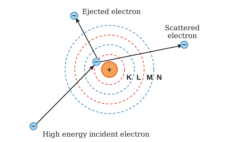
Ionizing radiations have enough energy to remove electrons from atoms, producing ions. X-rays, gamma rays and positrons are examples. X-rays and gamma rays interact with matter through the photoelectric effect and Compton scattering, and positrons are emitted from radioactive isotopes.

Non-Ionizing Radiations

Non-ionizing radiations do not have enough energy to remove electrons from atoms. Ultrasound waves and radio waves are examples; they are reflected, refracted or absorbed when they interact with matter.

Uses

X-rays image bones and internal organs.
Gamma rays and positrons are used in nuclear medicine, such as SPECT, PET-scans and cancer treatment, because of their high penetrating power.
Ultrasonic waves image soft tissues, monitor the fetus and study internal organs safely, without ionizing radiation.
Radio waves are used in MRI, interacting with atomic nuclei to image soft tissues without harmful radiation.

Ultrasound

Definition

Ultrasound is a widely used non-invasive medical imaging technique that shows internal body structures such as tissues, organs and the fetus during pregnancy.

Principle

It works on the piezoelectric effect, which is central to the ultrasound transducer. The transducer plays a dual role: it generates ultrasound waves and detects the signals reflected from inside the body.
LQ 1starts here — the piezoelectric effect, and how it allows transducers to both emit and detect ultrasound waves

Piezoelectric Effect

Definition

The piezoelectric effect is the generation of an electric charge by certain crystals when mechanical pressure is applied to them, and the change in their shape when an electric field is applied.

Name

The word piezoelectric comes from the Greek word piezein, meaning to press, and electric refers to the production of electricity.

Pioneer

Pierre Curie and Jacques Curie, two French brothers, in 1880.

Discovery

Experimenting with crystals such as quartz, tourmaline and Rochelle salt (sodium potassium tartrate), they found that crystals under mechanical pressure generate an electric charge, a strong connection between mechanical force and electrical response.

Cause

These crystals have no centre of symmetry. Inside them are small regions of positive and negative charge which are normally balanced.

Direct Piezoelectric Effect

When pressure is applied the balance is disturbed, the charges move slightly and an electric field appears on the surface.

Converse Piezoelectric Effect

When an electric field is applied to the crystal, it changes shape a little.
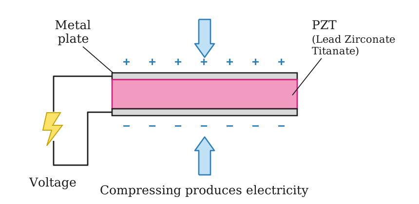
Fig. 20.1: Schematic diagram demonstrating piezoelectric effect

In a Transducer

The transducer generates ultrasound waves by the converse effect: an applied alternating voltage makes the crystal vibrate.
It detects the returning echoes by the direct effect: the echoes press the crystal, which produces a voltage.

Tidbit

The piezoelectric effect turns pressure into voltage and voltage into pressure. This is how ultrasound talks to the body.
LQ 1ends here

Short Questions

SQ 20.1.1

Distinguish between ionizing and non-ionizing radiations, with one example of each.

Ionizing radiations

They have enough energy to remove electrons from atoms, producing ions, such as X-rays and gamma rays.

Non-ionizing radiations

They cannot remove electrons from atoms, such as ultrasound and radio waves.
SQ 20.1.2

Why do piezoelectric crystals produce a voltage when pressed?

No centre of symmetry

Their positive and negative charge regions are normally balanced. Pressure disturbs the balance, the charges shift slightly and an electric field appears on the surface.
SQ 20.1.3

Distinguish between the direct and the converse piezoelectric effect.

Direct effect

Pressure on the crystal produces an electric field on its surface.

Converse effect

An electric field applied to the crystal makes it change shape.
SQ 20.1.4

Name three crystals with which the Curie brothers found the piezoelectric effect.

Crystals

Quartz, tourmaline and Rochelle salt (sodium potassium tartrate), in 1880.
MCQ 20.1.1

Medical physics is mainly used in:

Adiagnosis and treatment of diseasesBgenerating electricity
Cweather forecastingDspace travel
Solution:It applies physics to medicine.
MCQ 20.1.2

Which of these is a non-ionizing radiation?

AX-raysBultrasound
Cgamma raysDpositrons
Solution:It cannot remove electrons from atoms.
MCQ 20.1.3

MRI uses:

AX-raysBgamma rays
CultrasoundDradio waves
Solution:They interact with atomic nuclei to image soft tissues.
MCQ 20.1.4

The piezoelectric effect was discovered in:

A1895B1932
C1880D1970
Solution:By Pierre and Jacques Curie.
MCQ 20.1.5

The word piezein means:

Ato shineBto cut
Cto heatDto press
Solution:Pressure produces electricity.
MCQ 20.1.6

A transducer generates ultrasound by the:

Adirect piezoelectric effectBphotoelectric effect
Cconverse piezoelectric effectDDoppler effect
Solution:An alternating voltage makes the crystal vibrate.
MCQ 20.1.7

PZT stands for:

Apotassium zinc tartrateBlead zirconate titanate
Cplatinum zirconium titaniumDphosphorus zinc tungsten
Solution:It is the crystal of Fig. 20.1.
Topic 20.2

Diagnostic Use of Ultrasound in Medical Imaging

LQ 2starts here — how an ultrasound image is created using echoes and reflected sound waves

Ultrasound Imaging

Definition

Ultrasound imaging sends high frequency sound waves, typically 1 MHz to 15 MHz, into the body with a transducer and builds an image from the echoes reflected at the boundaries between tissues, in real time.
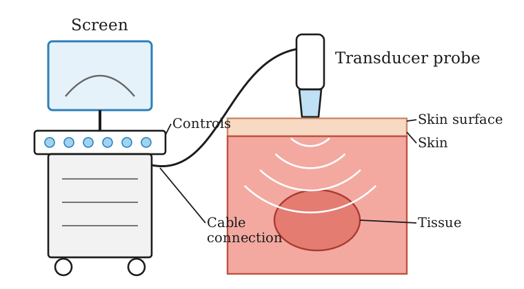
Fig. 20.2: Ultrasound machine

Advantage

Ultrasound does not use ionizing radiation, so it is a safe, non-invasive and repeatable imaging method.

Working

The piezoelectric transducer emits pulses of ultrasound waves, which travel at different speeds in different tissues.
At a boundary between two tissues of different acoustic impedance, determined by tissue density and sound speed, part of the wave is reflected.
The transducer switches to receive mode and detects the reflected waves by the direct piezoelectric effect.
The echoes are converted into electrical signals and processed by the machine.
A computer calculates the time delay and intensity of the returning echoes and constructs a visual image.

Brightness

Areas with stronger reflections appear brighter in the image, revealing the internal structures of organs and tissues.

Do You Know

The crystals in an ultrasound probe are smaller than a grain of rice, but they can both send and detect sound waves.
LQ 2ends here

Factors Affecting Image Quality

Acoustic Impedance Difference

A higher difference between two tissues, such as soft tissue and bone, causes stronger echoes and clear boundaries in the image.

Frequency of Ultrasound

Higher frequency ultrasound gives better resolution but less penetration; lower frequency waves penetrate deeper but give lower image clarity.

Use of Gel

A special gel between the transducer and the skin removes air gaps and improves the transmission of the sound waves.

Tidbit

Ultrasound can even measure the speed of blood flow using the Doppler effect, by detecting shifts in sound frequency.

Applications

Uses

Obstetrics: monitoring fetal growth, estimating gestational age and identifying abnormalities during pregnancy.
Abdomen: examining the liver, kidneys, gallbladder, pancreas and bladder.
Cardiology: echocardiography shows the heart’s structure and function in real time.
Urology: examining the prostate, testicles and urinary system.
Musculoskeletal system: detecting ligament injuries, joint inflammation and other soft tissue conditions.

Short Questions

SQ 20.2.1

Why is a gel applied between the ultrasound transducer and the skin?

Removing air gaps

The gel removes the air gaps between probe and skin, so the sound waves pass into the body instead of being reflected at the air.
SQ 20.2.2

How does the frequency of ultrasound affect the image?

Resolution against depth

Higher frequency gives better resolution but less penetration; lower frequency penetrates deeper but gives a less clear image.
SQ 20.2.3

Why do some regions of an ultrasound image appear brighter?

Stronger echoes

Areas that reflect the sound more strongly, at boundaries of large impedance difference, appear brighter.
SQ 20.2.4

What is the advantage of ultrasound over X-ray imaging?

No ionizing radiation

Ultrasound uses sound waves, so it is safe, non-invasive and repeatable, and is used to watch the fetus during pregnancy.
MCQ 20.2.1

The usual frequency range of medical ultrasound is:

A1 MHz to 15 MHzB20 Hz to 20 kHz
C1 kHz to 15 kHzD1 GHz to 15 GHz
Solution:Well above the audible range.
MCQ 20.2.2

Acoustic impedance is determined by:

Atissue density and sound speedBtissue temperature
Cthe colour of tissueDthe voltage of the probe
Solution:A difference in it at a boundary reflects the wave.
MCQ 20.2.3

Higher frequency ultrasound gives:

Aworse resolution and more penetrationBno change in the image
Cbetter resolution but less penetrationDionizing radiation
Solution:Lower frequency penetrates deeper.
MCQ 20.2.4

The speed of blood flow is measured with ultrasound using the:

Aphotoelectric effectBCompton effect
Cpiezoelectric effect onlyDDoppler effect
Solution:It detects the shift in sound frequency.
MCQ 20.2.5

Echocardiography is ultrasound imaging of the:

AkidneysBheart
CbrainDbones
Solution:It shows the heart’s structure and function in real time.
Topic 20.3

X-Rays

X-Rays

Definition

X-rays are high energy photons emitted when the inner shell electrons of heavy atoms make transitions back to their normal energy levels.

Light Elements

Transitions of electrons in hydrogen or other light elements emit spectral lines in the infrared, visible or ultraviolet region, because the differences between their energy levels are small.

Heavy Atoms

In heavy atoms the electrons are arranged in concentric shells K, L, M, N, O, with the K-shell closest to the nucleus. The inner shell electrons are tightly bound, so a large amount of energy is needed to knock them out of their normal levels.
Fig. 20.3: Concentric energy shells

Emission

When the atom returns to its normal state, photons of large energy are emitted, so inner shell transitions in heavy atoms give rise to X-rays.

Electromagnetic Spectrum

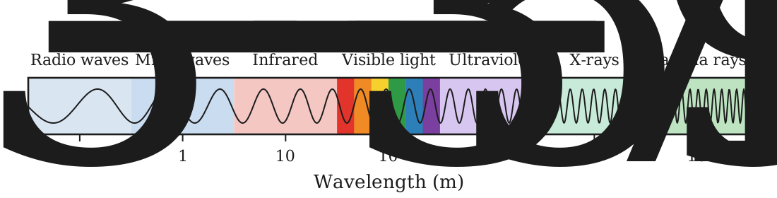
X-rays lie between ultraviolet and gamma rays, with wavelengths of about $10^{-9}$ m to $10^{-11}$ m.
Fig. 20.4: Electromagnetic spectrum
LQ 6starts here — inner shell transitions, and characteristic and continuous X-rays

Production of X-Rays

X-Ray Tube

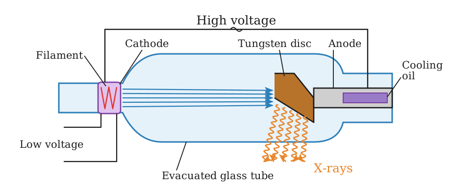
X-rays are produced in a high vacuum tube called the X-ray tube. The cathode C, heated by the filament F, emits electrons which are accelerated towards the anode T by the potential difference $V$.
Fig. 20.5: Schematic arrangement for producing X-rays

Energy of the Electrons

The maximum kinetic energy with which an electron strikes the target is
$$(K.E.)_{max}=Ve$$

Knocking Out a K Electron

These fast electrons strike a target of tungsten or another heavy element. In a collision an electron of an innermost shell, K or L, may be knocked out, leaving a vacancy or hole in that shell.

Characteristic X-Rays

Definition

The X-rays emitted in inner shell transitions are called characteristic X-rays, because their energies depend upon the type of target material.

K-alpha X-Ray

An electron from the L-shell jumps into the hole in the K-shell and emits a photon of energy $hf$, called the $K_{\alpha}$ X-ray.
$$hf_{K\alpha}=E_L-E_K$$

K-beta X-Ray

An electron from the M-shell may instead jump into the hole in the K-shell, emitting the $K_{\beta}$ X-ray.
$$hf_{K\beta}=E_M-E_K$$

More X-Rays

The holes created in the L and M-shells are filled by electrons from higher energy states, creating more X-rays. The characteristic X-rays appear as discrete lines on a continuous spectrum.

Use

The study of characteristic X-ray spectra has played a very important role in the study of atomic structure and the periodic table of elements.

The Continuous X-Ray Spectrum

Definition

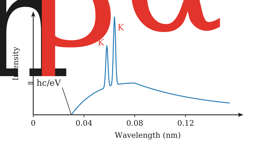
The continuous X-ray spectrum is the radiation emitted by the fast electrons as they are suddenly decelerated in the target, an effect known as bremsstrahlung or braking radiation.

Cause

An accelerating charge emits electromagnetic radiation. The rate of deceleration is so large that the emitted radiation has a short wavelength, so the bremsstrahlung lies in the X-ray region.

Shortest Wavelength

An electron that loses all its kinetic energy in the first collision gives a single photon of the maximum frequency.
$$(K.E.)_{max}=hf_{max}$$
This frequency corresponds to the shortest wavelength.
$$\lambda_{min}=\frac{hc}{eV}$$
Fig. 20.6: Continuous X-ray spectrum

Longer Wavelengths

Other electrons do not lose all their energy in the first collision and suffer a number of collisions before coming to rest. They give photons of smaller energy, X-rays of longer wavelength, so a continuous spectrum is obtained.
LQ 6ends here
LQ 3starts here — how attenuation of X-rays by different tissues creates contrast in the image

X-Ray Imaging

Attenuation

As X-rays travel through the body they lose energy because they interact with atoms. This process is called attenuation. X-rays ionize atoms, knocking out electrons, which is how their energy is transferred.

Exponential Decrease

The intensity of the X-ray beam decreases gradually as it passes through a material, following an exponential pattern.

Image Intensifiers

Image intensifiers are used in real-time X-ray imaging. They brighten the image, which makes it easier to see and reduces the radiation exposure.

Contrast

Definition

Contrast in X-ray images means how clearly we can tell different tissues apart.

Depends on Energy

Hard X-rays, of high energy, are used for bone imaging.
Soft X-rays, of low energy, are better for soft tissues such as the breast.

Contrast Media

Special substances such as iodine or barium, which absorb X-rays well, are injected or swallowed to temporarily enhance the visibility of internal structures, organs and blood vessels.
LQ 3ends here

Short Questions

SQ 20.3.1

Why do heavy atoms emit X-rays while light atoms emit visible or UV light?

Energy gaps

The inner shell electrons of heavy atoms are tightly bound, so the energy differences of their transitions are large and the photons are X-rays. In light atoms the levels are close together.
SQ 20.3.2

What is bremsstrahlung?

Braking radiation

The X-radiation emitted when fast electrons are suddenly decelerated in the target. It gives the continuous X-ray spectrum.
SQ 20.3.3

On what does the shortest wavelength of the continuous X-ray spectrum depend?

Accelerating voltage

An electron losing all its energy in one collision gives the shortest wavelength.
$$\lambda_{min}=\frac{hc}{eV}$$
It depends only on the tube voltage $V$.
SQ 20.3.4

What is attenuation of X-rays?

Attenuation

The loss of energy of X-rays as they pass through the body and interact with atoms. The beam intensity falls exponentially with distance.
SQ 20.3.5

Distinguish between hard and soft X-rays in imaging.

Hard X-rays

High energy X-rays, used for bone imaging.

Soft X-rays

Low energy X-rays, better for soft tissues such as the breast.
SQ 20.3.6

What are contrast media? Give two examples.

Contrast media

Substances that absorb X-rays well, injected or swallowed to make internal organs and blood vessels show up. Iodine and barium are examples.
MCQ 20.3.1

The shell closest to the nucleus is the:

AL-shellBM-shell
CK-shellDN-shell
Solution:Then L, M, N and O.
MCQ 20.3.2

An electron accelerated through 30 kV strikes the target with a maximum K.E. of:

A$4.8\times10^{-24}$ JB$1.9\times10^{-14}$ J
C$3.0\times10^{4}$ JD$4.8\times10^{-15}$ J
Solution:(K.E.)max = Ve = 30000 x 1.6e-19 = 4.8e-15 J.
MCQ 20.3.3

The shortest X-ray wavelength from a tube at 41.4 kV is about:

A0.3 nmB0.03 nm
C3 nmD0.003 nm
Solution:λ = hc/eV = 1.99e-25/(1.6e-19 x 41400) = 3.0e-11 m.
MCQ 20.3.4

If the tube voltage is doubled, the shortest X-ray wavelength:

Ais halvedBis doubled
Cstays the sameDis four times
Solution:λmin = hc/eV.
MCQ 20.3.5

The continuous X-ray spectrum is due to:

Ainner shell transitionsBbremsstrahlung
Cnuclear decayDannihilation
Solution:Braking radiation of decelerated electrons.
MCQ 20.3.6

The target of an X-ray tube is usually made of:

AcopperBaluminium
CcarbonDtungsten
Solution:A heavy element.
MCQ 20.3.7

Which substance is swallowed as an X-ray contrast medium?

AbariumBsodium
CcarbonDhelium
Solution:Iodine is injected; both absorb X-rays well.
Topic 20.4

Computed Tomography Scanning (CT-Scan)

LQ 4starts here — CT compared with conventional X-ray imaging in image clarity and diagnostic value

CT-Scan

Definition

Computed tomography, the CT-scan, is a medical imaging technique that combines X-ray technology with computer processing to generate high resolution cross-sectional and 3D images of internal body structures.

Use

It is especially useful for diagnosing diseases, detecting internal injuries and guiding medical treatment.

Problem with an Ordinary X-Ray

An ordinary X-ray shows a flat 2D image in which bones and organs overlap each other, so individual structures are hard to see.

Working

A CT-scanner takes many X-ray images of the same body section from different angles.
A computer combines them into a detailed cross-sectional slice of the body.
The scanner moves slightly along the body and repeats the process for the next slice.
All the slices are combined by the computer into a 3D image of the internal structure.

The Scanner

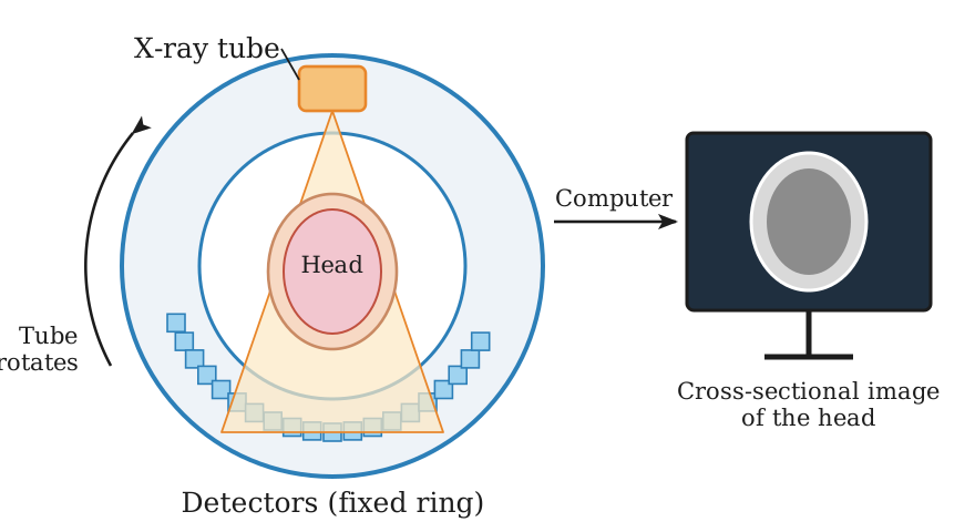
The X-ray tube rotates around the patient while the detectors stay in place, collecting complete information from all sides.
Fig. 20.7: A boy 3D head cross-section CAT-scan

Name

Computed: a computer processes the data.
Tomography: from the Greek word tomos, meaning slice.
Axial: the scan is taken along the body’s axis.

CAT-Scan

The technique is called computed axial tomography, CAT-scan, because a computer controls the scanning, collects the data and converts it into images while the X-ray tube rotates around the body.

Do You Know

The CT-scanner was invented by Sir Godfrey Hounsfield (England) and Allan Cormack (South Africa) in the early 1970s, who shared the Nobel Prize in Physiology or Medicine in 1979.
CT-scans create hundreds of slices in seconds, like peeling a digital apple layer by layer, and can detect strokes within minutes of onset.

Advantages of CT-Scan

Over X-Rays

A single X-ray image is quick to produce but gives only a flat 2D view. A CT-scan produces 3D images showing the size, shape and exact location of organs, bones and other structures.

Similar Densities

It distinguishes between tissues even when their densities are very similar. It can clearly show the position and size of a tumour, which helps doctors target it accurately with high energy X-rays or gamma rays.

Risk

A CT-scan involves exposure to X-rays, a form of ionizing radiation. Its dose is comparable to, or a few times greater than, one year of natural background radiation, or roughly that of four long-distance flights, so doctors take special precautions with pregnant women and patients with existing health concerns.

Tidbit

CT-scanners take X-ray images from different angles to reconstruct a 2D cross-section; combining the slices makes a 3D model, like stacking the layers of a cake.
LQ 4ends here

Short Questions

SQ 20.4.1

Where does the word tomography come from?

Tomos

From the Greek word tomos, meaning slice; a CT-scan builds the body up slice by slice.
SQ 20.4.2

Why is a CT-scan also called a CAT-scan?

Computed axial tomography

A computer controls the scan and the images are taken along the body’s axis as the X-ray tube rotates round the patient.
SQ 20.4.3

Who invented the CT-scanner?

Inventors

Sir Godfrey Hounsfield and Allan Cormack, in the early 1970s; they shared the Nobel Prize in Physiology or Medicine in 1979.
SQ 20.4.4

Why must special precautions be taken with CT-scans?

Ionizing radiation

A CT-scan exposes the patient to X-rays, about one year of background radiation or four long-distance flights, so pregnant women and sick patients need special care.
MCQ 20.4.1

In a CT-scanner the part that rotates around the patient is the:

AdetectorsBpatient table
CX-ray tubeDmonitor
Solution:The detectors stay in place.
MCQ 20.4.2

A CT-scan makes a 3D image by combining:

Across-sectional slicesBechoes of sound
Cgamma-ray pairsDradio signals
Solution:Each slice is made from many X-ray views.
MCQ 20.4.3

The CT-scanner was invented in the:

Aearly 1900sB1880s
Cearly 1970sD2010s
Solution:By Hounsfield and Cormack.
MCQ 20.4.4

The radiation dose of a CT-scan is roughly equal to:

Aone dental X-rayBzero
Cone minute of sunlightDfour long-distance flights
Solution:Or about one year of natural background radiation.
Topic 20.5

Positron Emission Tomography Scanning (PET-Scan)

LQ 5starts here — PET, and how it is used to scan malignant tissues

PET-Scan

Definition

Positron emission tomography, the PET-scan, is a non-invasive technique that images metabolic and biochemical activity at the cellular or molecular level, using a positron-emitting tracer inside the body.

Imaging with Radiation

As photons pass through tissue they may be absorbed or scattered, and the pattern of energy deposition and attenuation produces measurable signals at a detector.

Nuclear Medicine

In nuclear medicine the radiation originates inside the patient from an administered radiopharmaceutical, so the signal reflects physiological function rather than only anatomy.

SPECT

A gamma-emitting tracer is introduced into the body and a rotating gamma camera records many projection views, which are reconstructed into cross-sectional images of the tracer, giving 3D information about perfusion and organ function.

PET

PET extends this idea with positron-emitting tracers. The positron travels a short distance in tissue and annihilates with an electron, producing two gamma photons in nearly opposite directions; detecting them in coincidence locates the event and builds a map of tracer uptake.

Uses

Early cancer detection and staging.
Evaluating brain function.
Identifying cardiac abnormalities before clear anatomical changes appear.

Working of PET-Scan

Tracer

A small amount of radioactive tracer is injected into a vein. The blood carries it and it builds up more in very active tissues, such as many cancers and the working parts of the brain and heart.

Annihilation

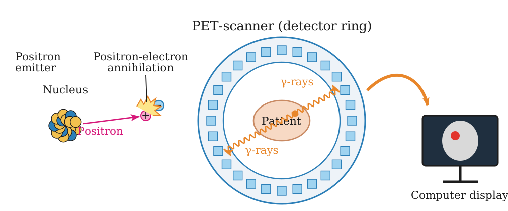
The unstable nuclei of the tracer decay and emit positrons. Each positron travels only a short distance before meeting an electron in nearby tissue; the two annihilate and their mass becomes two gamma-ray photons moving in opposite directions.
Fig. 20.8: Schematic diagram of PET-scanning

Detection

Detectors arranged in a ring around the patient detect these photon pairs and send the signals to a computer, which constructs images showing where the tracer has collected.

Image

Brighter areas on a PET image are regions of higher tracer concentration and therefore higher tissue activity, which is how malignant tissue is found.
LQ 5ends here

Short Questions

SQ 20.5.1

Why does a PET tracer collect more in cancerous tissue?

Active tissue

The tracer is carried by the blood and builds up where tissue is most active, and many cancers are very active.
SQ 20.5.2

What is SPECT?

SPECT

A technique in which a gamma-emitting tracer is introduced into the body and a rotating gamma camera records views that are reconstructed into cross-sectional images of the tracer.
SQ 20.5.3

How does PET differ from X-ray imaging?

Function, not anatomy

In PET the radiation comes from inside the patient, so the image shows metabolic activity; an X-ray image shows the structure the beam passes through.
MCQ 20.5.1

In a PET-scan the tracer emits:

AelectronsBpositrons
Calpha particlesDneutrons
Solution:Its unstable nuclei decay by positron emission.
MCQ 20.5.2

The two gamma photons of an annihilation move:

Ain the same directionBin opposite directions
Cat right anglesDin random directions
Solution:Momentum is conserved.
MCQ 20.5.3

Each gamma photon from electron-positron annihilation at rest has an energy of:

A0.51 MeVB1.02 MeV
C0.255 MeVD2.04 MeV
Solution:The pair’s 1.02 MeV is shared by two photons.
MCQ 20.5.4

Brighter areas on a PET image show:

Adenser boneBair spaces
Cstronger echoesDhigher tissue activity
Solution:More tracer has collected there.
MCQ 20.5.5

PET detectors are arranged:

Ain a single lineBinside the patient
Cin a ring around the patientDon the computer
Solution:They catch the photon pairs from all sides.

Chapter Exercise

SQ 20.1

Which type of radiation is produced during a PET-scan?

Gamma rays

A positron from the tracer annihilates with an electron, and their mass becomes two gamma-ray photons moving in opposite directions.
SQ 20.2

Which physical effect enables ultrasound transducers to produce sound waves?

Piezoelectric effect

By the converse piezoelectric effect an alternating voltage applied to the crystal makes it vibrate, producing the ultrasound waves. The direct effect detects the returning echoes.
SQ 20.3

Mention one area of the body commonly examined using ultrasound.

Abdomen

Ultrasound examines the abdomen, the liver, kidneys, gallbladder, pancreas and bladder. It also monitors the fetus during pregnancy.
SQ 20.4

What causes contrast in X-ray images of different body tissues?

Different attenuation

Tissues of different density and thickness absorb X-rays differently. Bone absorbs more than soft tissue, so the transmitted intensity, and the image, differ from one tissue to the next.
SQ 20.5

What occurs when ultrasound hits the boundary between two tissues?

Part of the wave is reflected.

Acoustic impedance

At a boundary between tissues of different acoustic impedance part of the wave is reflected as an echo and the rest is transmitted. The greater the difference, the stronger the echo.
SQ 20.6

How does CT-scan improve imaging? Explain briefly.

Slices instead of a shadow

A CT-scanner takes many X-ray images of one section from different angles, and a computer combines them into a cross-sectional slice; the slices are then combined into a 3D image. Organs no longer overlap as in a flat X-ray, so their size, shape and position are seen clearly.
SQ 20.7

What do you know about the terms kα and kβ X-rays.

K-alpha X-ray

Emitted when an electron from the L-shell fills a hole in the K-shell.
$$hf_{K\alpha}=E_L-E_K$$

K-beta X-ray

Emitted when an electron from the M-shell fills the K-shell hole, so it has higher energy.
$$hf_{K\beta}=E_M-E_K$$
CRQ 20.1

What does a time difference in detecting gamma rays at two detectors indicate in a PET scan?

The annihilation did not occur midway between the two detectors.

Locating the event

The two gamma photons leave the annihilation point together at the speed of light. If one reaches its detector earlier, the event was closer to that detector; the time difference fixes how far along the line between the detectors the annihilation took place, which sharpens the image.
CRQ 20.2

Why is ultrasound not effective for imaging air-filled organs like the lungs? Explain.

Almost total reflection

The acoustic impedance of air is very different from that of soft tissue, so at a tissue-air boundary nearly all the ultrasound is reflected. Little energy enters the air-filled lung, and air also absorbs ultrasound strongly, so no useful echoes come back from inside it. This is also why gel is used between the probe and the skin.
CRQ 20.3

What happens when the X-ray contrast between two tissues is too low? Suggest a solution.

The two tissues cannot be told apart in the image.

Solutions

Use a contrast medium, such as iodine injected or barium swallowed, which absorbs X-rays strongly and highlights the organ or blood vessels.
Use soft (low energy) X-rays for soft tissues, which differ more in their absorption.
Use a CT-scan, which can distinguish tissues of very similar density.
CRQ 20.4

How does Computed Tomography scanning reduce the overlapping of organs seen in plain X-rays? Explain.

Imaging one slice at a time

A plain X-ray projects the whole body thickness onto one flat film, so organs lie on top of each other. In CT the X-ray tube rotates round the patient and takes many images of one thin section from different angles; the computer reconstructs that slice alone. Repeating this along the body gives separate slices, combined into a 3D image, so no organ hides another.
CRQ 20.5

What could be a possible medical cause of uneven tracer concentration seen in a PET brain scan?

Uneven brain activity

The tracer collects where tissue is most active. A region of high concentration may be a tumour, whose cells are very active, or a seizure focus; a region of low concentration may show damaged or less active tissue, as after a stroke or in dementia.
CRQ 20.6

What is the purpose of lead collimators in PET gamma-ray detectors? Explain.

Accepting rays from one direction

Lead absorbs gamma rays strongly. Collimators of lead in front of the detectors let in only the gamma rays travelling along the required direction and block scattered and stray radiation, so each detected photon pair points back correctly to its annihilation site and the image is sharp.
MCQ 20.1

What kind of radiation is detected in a PET scan after positron annihilation?

ABeta particlesBGamma rays
CX-raysDAlpha particles
Solution:The positron and electron annihilate into two gamma-ray photons moving in opposite directions.
MCQ 20.2

The role of coincidence detection in PET-scanning is:

Ato control the temperature of the scannerBto detect gamma rays arriving simultaneously
Cto block unwanted radiationDto generate X-rays
Solution:The two photons of one annihilation reach opposite detectors together, which locates the event.
MCQ 20.3

Which principle allows ultrasound transducers to emit and detect sound waves?

APiezoelectric effectBDoppler effect
CPhotoelectric effectDThermoelectric effect
Solution:The converse effect emits the waves and the direct effect detects the echoes.
MCQ 20.4

The body part which commonly examined using ultrasound imaging is:

AmusclesBbones
CabdomenDteeth
Solution:The liver, kidneys, gallbladder, pancreas and bladder are examined with ultrasound.
MCQ 20.5

In X-ray imaging, what does "contrast" mainly depend on?

ADensity and thickness of tissuesBHeartbeat rate
CDistance from the detectorDSurface temperature of the body
Solution:Denser, thicker tissue attenuates more X-rays, so it shows apart from its neighbours.
MCQ 20.6

What advantage does a CT-scan have over a standard X-ray image?

ALess radiation exposureBSimpler equipment
CProduces 3D images from 2D slicesDUses sound waves instead of X-rays
Solution:A computer combines many cross-sectional slices into a 3D image.
MCQ 20.7

The wave used in ultrasound imaging is:

Alongitudinal sound waveBtransverse sound wave
Celectromagnetic waveDionizing radiation wave
Solution:Ultrasound is a sound wave, and sound in tissue travels as a longitudinal wave.
MCQ 20.8

Characteristic X-rays are produced due to:

Anuclear reactionsBinner shell electronic transitions
Couter shell transitionsDion collisions
Solution:An L or M electron falling into a K-shell hole emits the K-alpha or K-beta line.
MCQ 20.9

What does “CT” stand for in medical imaging?

ACombined TomographyBComputed Telemetry
CComputed TomographyDCentralized Therapy
Solution:Computed, as a computer processes the data; tomography, from tomos, a slice.

Self Test

Seventeen questions across the whole chapter. Work through them without the notes, then check the answers keyed at the foot of the page.

Q 1

Ultrasound, X-rays and PET all help doctors to:

Acure diseases by heatingBmeasure blood pressure
Csee inside the body without surgeryDreplace organs
Q 2

Gamma rays and positrons are used in nuclear medicine because of their:

Alow energyBlong wavelength
Caudible frequencyDhigh penetrating power
Q 3

A piezoelectric crystal has:

Afree electrons throughoutBno centre of symmetry
Ca magnetic field inside itDzero density
Q 4

A transducer detects the returning echoes by the:

Adirect piezoelectric effectBconverse piezoelectric effect
Cphotoelectric effectDCompton effect
Q 5

In an ultrasound image the computer uses the echoes’:

Acolour and chargeBmass and charge
Ctime delay and intensityDtemperature only
Q 6

Ultrasound is reflected most strongly at a boundary between:

Atwo similar soft tissuesBsoft tissue and bone
Ctwo layers of the same tissueDblood and blood
Q 7

In obstetrics ultrasound is used to:

Aimage the lungsBtreat cancer
Cmeasure bone density by X-raysDmonitor fetal growth
Q 8

Characteristic X-rays have energies that depend upon the:

Atarget materialBtube current
Csize of the tubeDtemperature of the room
Q 9

A K-alpha X-ray is emitted when an electron falls into the K-shell from the:

AL-shellBM-shell
CN-shellDnucleus
Q 10

The maximum K.E. of the electrons striking an X-ray target is:

Ahc/λBVe
Cmc²DVe/2
Q 11

The intensity of an X-ray beam passing through tissue decreases:

AlinearlyBnot at all
CexponentiallyDin steps
Q 12

Real-time X-ray imaging uses image intensifiers to:

Aproduce sound wavesBcool the target
Cemit positronsDbrighten the image and reduce exposure
Q 13

In a CT-scanner the detectors:

Arotate with the tubeBmove along the body
Cstay in placeDare inside the patient
Q 14

A CT-scan can show a tumour’s:

AtemperatureBposition and size
Celectric chargeDsound speed only
Q 15

In nuclear medicine the radiation comes from:

Aan X-ray tubeBa transducer
Cthe sunDinside the patient
Q 16

A positron travels a short distance in tissue and then:

Aannihilates with an electronBsplits a nucleus
Cis absorbed by boneDbecomes a neutron
Q 17

PET is valuable for early cancer detection because it images:

Aonly bone structureBskin colour
Cmetabolic activityDblood temperature

Answers 1 (c)2 (d)3 (b)4 (a)5 (c)6 (b)7 (d)8 (a)9 (a)10 (b)11 (c)12 (d)13 (c)14 (b)15 (d)16 (a)17 (c)